41 basic animal cell diagram with labels
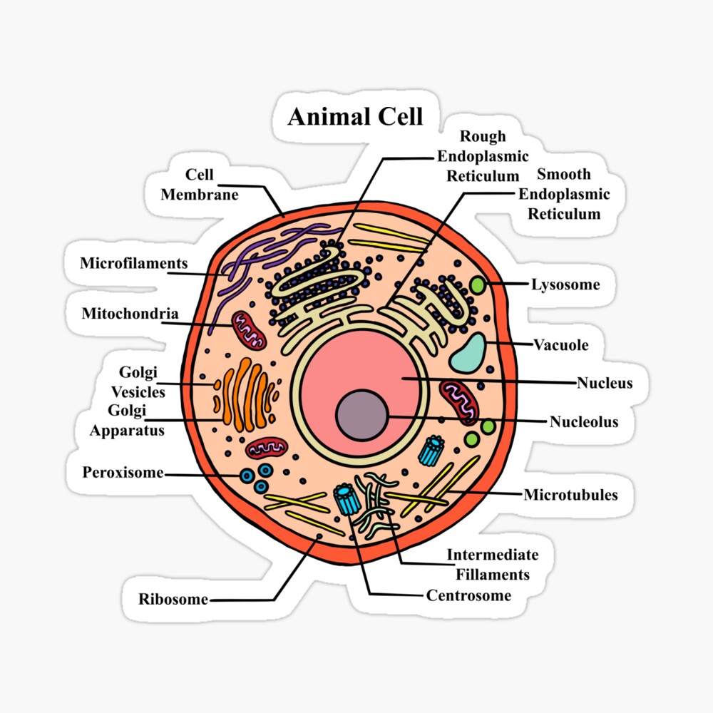
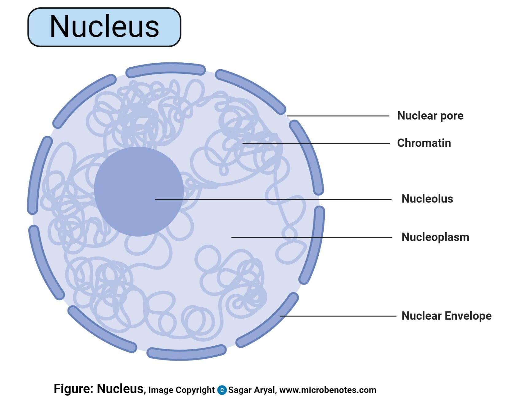
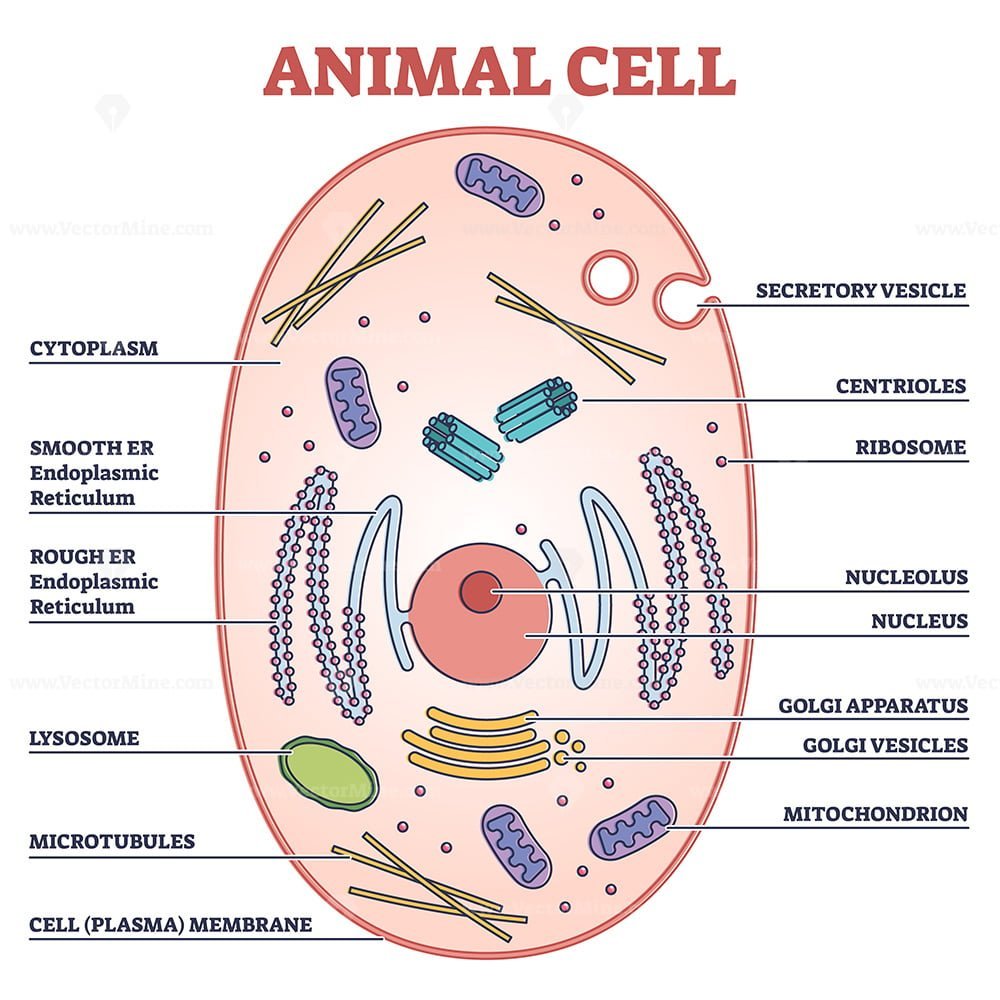
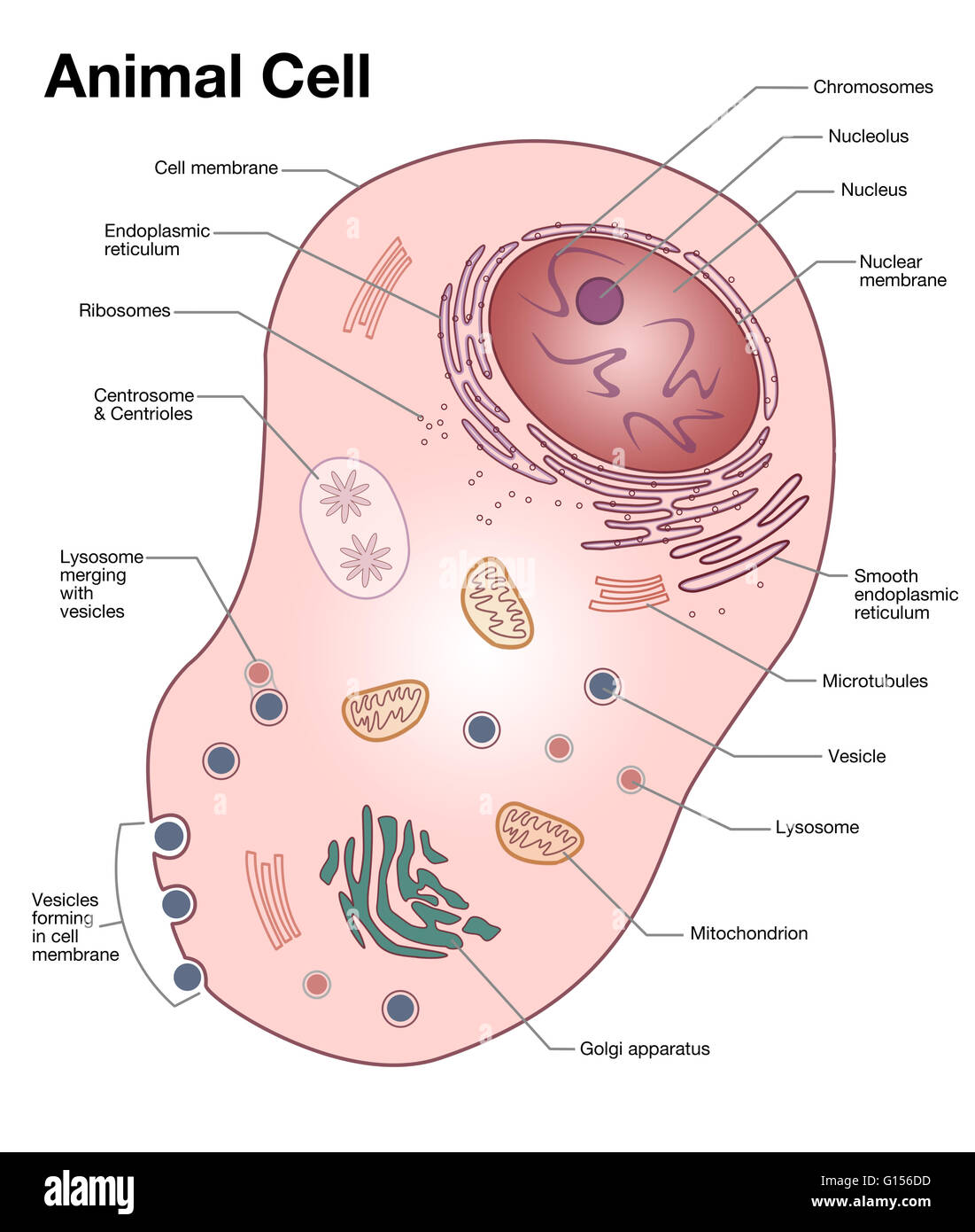
An Overview of Hyphae in Fungi, Their Function and Types. 10.01.2022 · Fungi have their cell wall made up of chitin. Their body is composed of long thread-like filaments or tubes known as hyphae . In singular form, this structure is referred to as a hypha. The Parts Of An Animal Cell | Science Trends Jessica McGregor. There are 13 main parts of an animal cell: cell membrane, nucleus, nucleolus, nuclear membrane, cytoplasm, endoplasmic reticulum, Golgi apparatus, ribosomes, mitochondria, centrioles, cytoskeleton, vacuoles, and vesicles. A cell is the smallest unit of life; cells tend to be 1 - 100 micrometers (μm) in diameter, and each ...
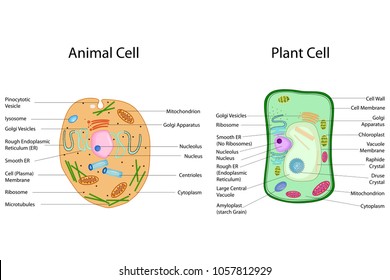
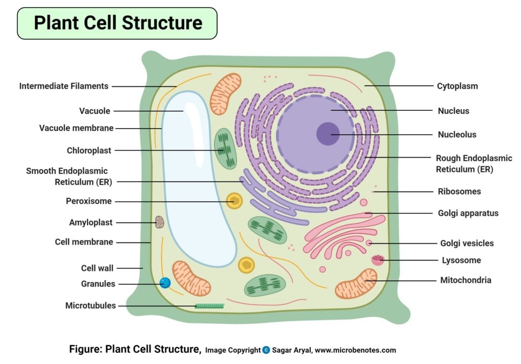
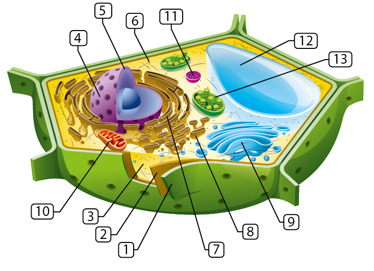
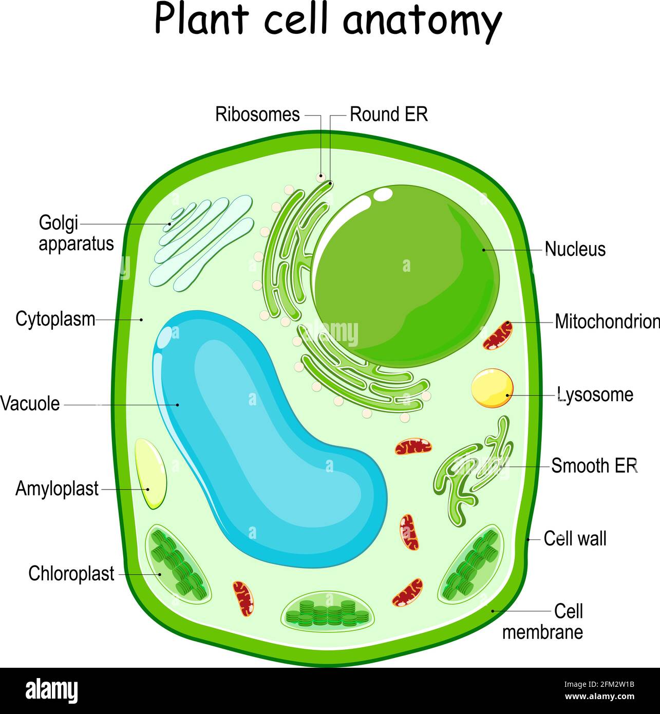
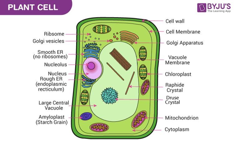
Plant and Animal Cell: Labeled Diagram, Structure, Function - Embibe Diagram of Plant and Animal Cell Fig: Plant Cell Fig: Animal Cell Learn Exam Concepts on Embibe Plant and Animal Cell Structures Both plant and animal cells have similar types of architecture. They are made up of cell boundaries, cytoplasm, nucleus and several cellular organelles. Different Plant and Animal Cell Parts Fig: Chloroplast

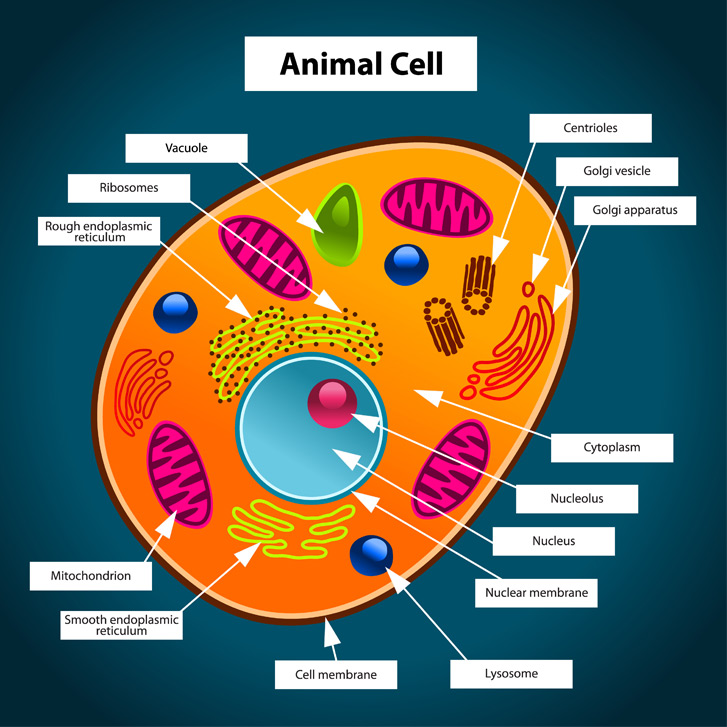
Basic animal cell diagram with labels
Interactive Bacteria Cell Model - CELLS alive Periplasmic Space: This cellular compartment is found only in those bacteria that have both an outer membrane and plasma membrane (e.g. Gram negative bacteria).In the space are enzymes and other proteins that help digest and move nutrients into the cell. Cell Wall: Composed of peptidoglycan (polysaccharides + protein), the cell wall maintains the overall shape of a … Animal and Plant Cell Worksheets - Super Teacher Worksheets This is a basic illustration of a plant cell with major parts labeled. Labels include nucleus, chloroplast, cytoplasm, membrane, cell wall, and vacuole, and mitochondrion. Use it as a poster in your classroom or have students glue it into their science notebooks. View PDF Plant Cell Vocabulary Cards Free Printable Plant and Animal Cells Worksheets 27.06.2022 · This animal cell diagram is labeled in cursive writing for your students who are learning about ... Plant Cell Parts Color Poster – This free plant cell parts poster is in full color to give a basic illustration of a plant cell including labels for the nucleus, chloroplast, cytoplasm , membrane, cell wall, vacuole, and mitochondrion. Learning About Plant Cells Diagram Printable …
Basic animal cell diagram with labels. Label the Animal Cell: Level 1 | Worksheet | Education.com In Label the Animal Cell: Level 1, students will use a word bank to label the parts of a cell in an animal cell diagram. To take the learning one step further, have students assign a color to each of the organelles and then color in the diagram. For a broader focus, use this worksheet in conjunction with the Label the Plant Cell: Level 1 worksheet. Wikipedia:Featured picture candidates/Cell membrane ... Edit 3 uploaded Standard zoom boxes tend to obscure the labels, but if there is a consensus to change to those, I'll give it another try. Dhatfield ( talk ) 20:29, 16 June 2008 (UTC) Reply [ reply ] Strong oppose There are only unsaturated tails on the phospholipids, for a reduced structure diagram of a cell membrane's lipid bilayer there should be one unsaturated and one saturated … Label Cell Parts | Plant & Animal Cell Activity | StoryboardThat Create a cell diagram with each part of plant and animal cells labeled. Include descriptions of what each organelle does. Click "Start Assignment". Find diagrams of a plant and an animal cell in the Science tab. Using arrows and Textables, label each part of the cell and describe its function. Animal Cells | Basic Biology Skin cells. The skin cells of animals mostly consist of keratinocytes and melanocytes - 'cyte' meaning cell. Keratinocytes make up around 90% of all skin cells and produce a protein called 'keratin'. The keratin in skin cells helps to make skin an effective layer of protection for the body. Keratin also makes hair and nails.
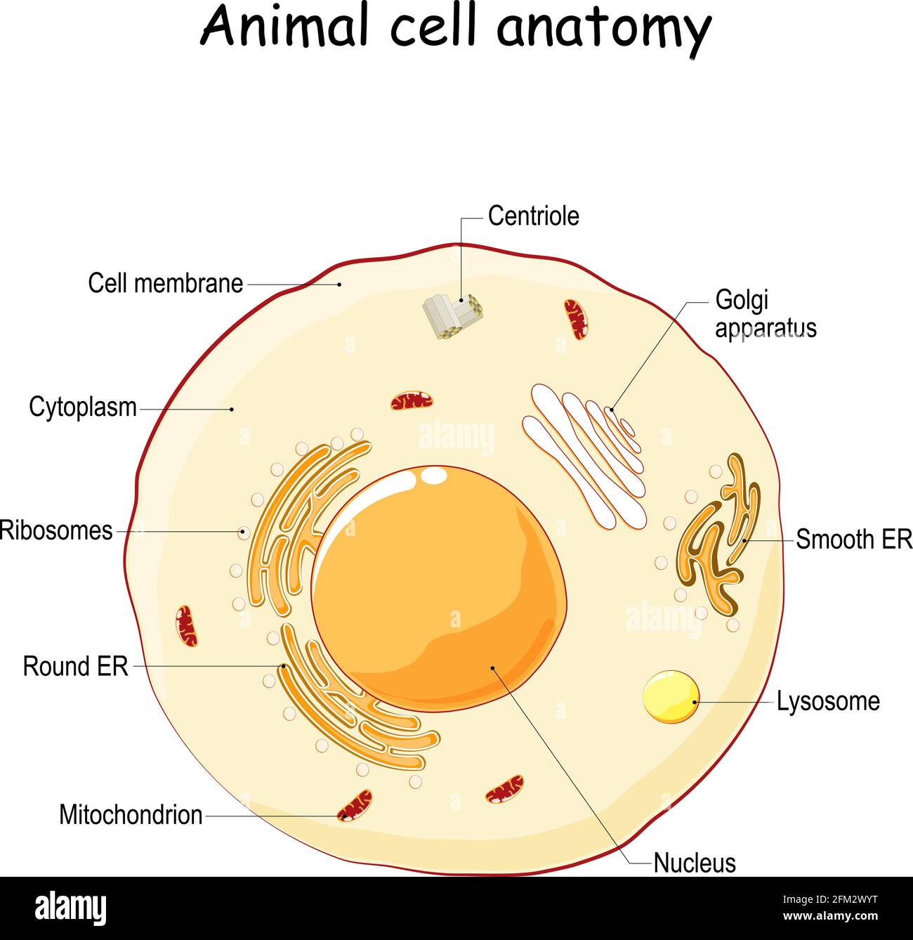
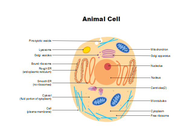
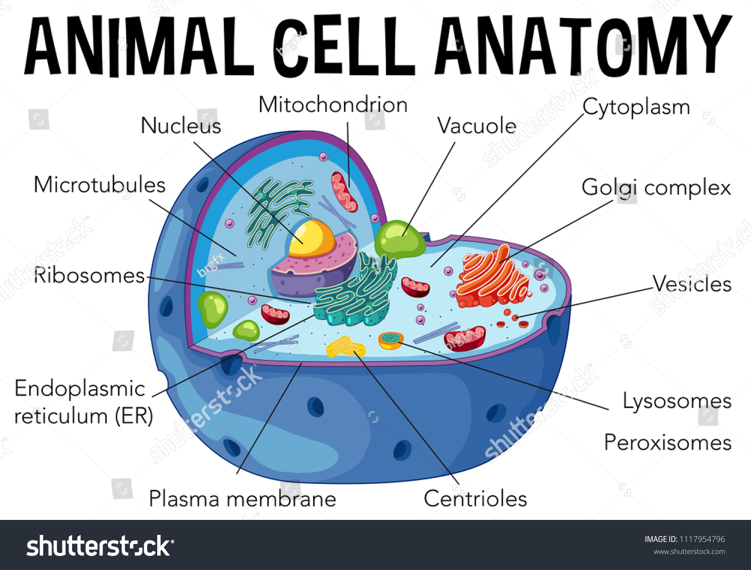
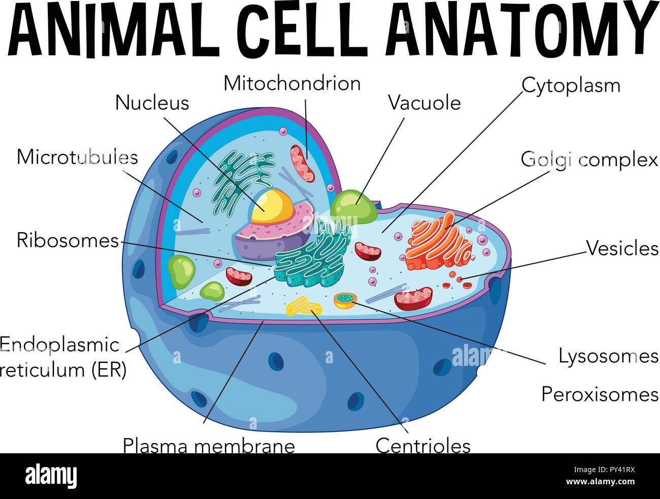
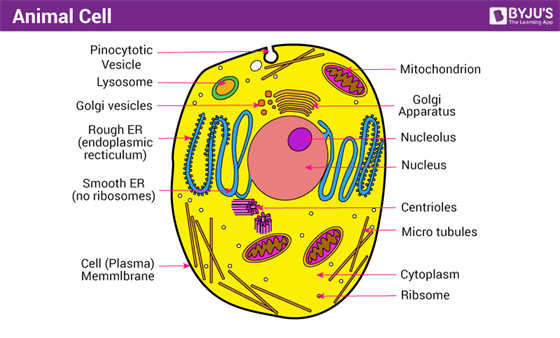
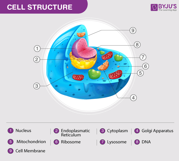
Junqueira's Basic Histology Text and Atlas, 14th Edition Junqueiras Basic Histology Text and Atlas 14th Edition Vet Books ir. by Zelle Peredas. Download Free PDF Download PDF Download Free PDF View PDF. Basic Histology. by MARIAN ESTRADAR. Download Free PDF Download PDF Download Free PDF View PDF. Color Atlas of Cytology, Histology, and Microscopic Anatomy. by Mina Ungureanu. Download Free PDF Download PDF … A Well-labelled Diagram Of Animal Cell With Explanation - BYJUS The animal cell diagram is widely asked in Class 10 and 12 examinations and is beneficial to understand the structure and functions of an animal. A brief explanation of the different parts of an animal cell along with a well-labelled diagram is mentioned below for reference. Also Read Different between Plant Cell and Animal Cell Plant Cells Vs. Animal Cells (With Diagrams) - Owlcation The most important structures of plant and animal cells are shown in the diagrams below, which provide a clear illustration of how much these cells have in common. The significant differences between plant and animal cells are also shown, and the diagrams are followed by more in-depth information. Diagram of an animal cell Doc Sonic Animal Cell Parts - Biology Wise The labeled diagram given below depicts the parts of an animal cell, which will help you in understanding the concept better. Labeled Animal Cell Diagram What Are the Various Parts of an Animal Cell? Cell Membrane: The cell membrane is the outermost part of the cell, which encloses all the other cell organelles.
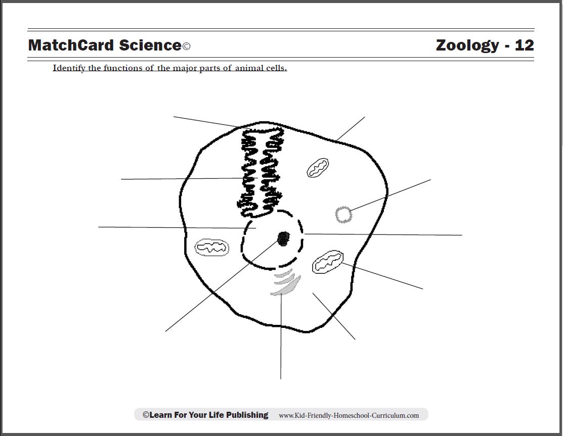
Animal Cell Anatomy & Diagram - Enchanted Learning Label the animal cell diagram, with a glossary of animal cell terms included. Answers. Make your own giant animal cell model using Jello and candies (and then eat it). Label the animal cell mitosis diagram. Answers. Label the axon, dendrites, cell body, nucleus, Schwann's cells, and nodes of Ranvier. Answers. Plant Cell Venn diagram - Wikipedia A Venn diagram is a widely used diagram style that shows the logical relation between sets, popularized by John Venn (1834–1923) in the 1880s. The diagrams are used to teach elementary set theory, and to illustrate simple set relationships in probability, logic, statistics, linguistics and computer science.A Venn diagram uses simple closed curves drawn on a plane to represent sets. Cell Worksheets | Plant and Animal Cells - Math Worksheets 4 Kids This collection of animal and plant cell worksheets strikes a balance between cognitive and psychomotor domains of learning and offers a conceptual grounding in cell biology. The worksheets recommended for students of grade 4 through grade 8 feature labeled animal and plant cell structure charts and cross-section charts, cell vocabulary with ... Animal Label Me! Printouts - EnchantedLearning.com Bird External Anatomy Printout. Label the bird's external anatomy on this printout. Answers. Birds and Bird Terms. Label the birds (and related terms), including: feather, wing, nest, egg, beak, penguin, pigeon, eagle, hummingbird, and swan. Answers. Birds in French. Label the birds (and related terms) in French.

Printable Animal Cell Diagram – Labeled, Unlabeled, and Blank
Animal cells - Cell structure - AQA - BBC Bitesize Animal cells have a basic structure. Below the basic structure is shown in the same animal cell, on the left viewed with the light microscope, and on the right with the transmission electron...
Labeled Animal Cell Diagram" Poster for Sale by BundaBear ...
animal cells without labels Animal Cell Labelling Activity | Basic Animal Cell Diagram. 15 Pictures about Animal Cell Labelling Activity | Basic Animal Cell Diagram : 30 Label Of Animal Cell, Animal cell - 3D - YouTube and also visual aid project for cells - Google Search | Science/Physics/Energy. Animal Cell Labelling Activity | Basic Animal Cell Diagram
Animal cell hi-res stock photography and images - Alamy
What Is An Animal Cell? Facts, Pictures & Info For Kids & Students. Most animal cells are between 10 and 20 micrometers across. A micrometer is one millionth of a meter, or one thousandth of a millimeter. In other words, most animal cells are very small! Although most animal cells are far too small to be seen without a microscope, some are much larger. The human egg cell, for example, is visible to the naked eye.
Animal Cell Diagram Labeled | EdrawMax Template
The Cell - ScienceQuiz.net Look at the diagram of an animal cell. Select correct statement from the following about animal cells. Select correct statement from the following about animal cells. A is the cell membrane and DNA is located inside B.

Plant Cell Diagram | Animal Cell Diagram | Plant and animal ...
Animal Cells: Labelled Diagram, Definitions, and Structure - Research Tweet Animal Cells: Labelled Diagram, Definitions, and Structure What is a Cell? In biology, cell is the smallest unit that can live on its own and that makes up all living organisms and the tissues of the body. A cell has three main parts: the cell membrane, the nucleus, and the cytoplasm. What is Animal Cell?
Draw a well labeled diagram of animal cell - Home Work Help ...
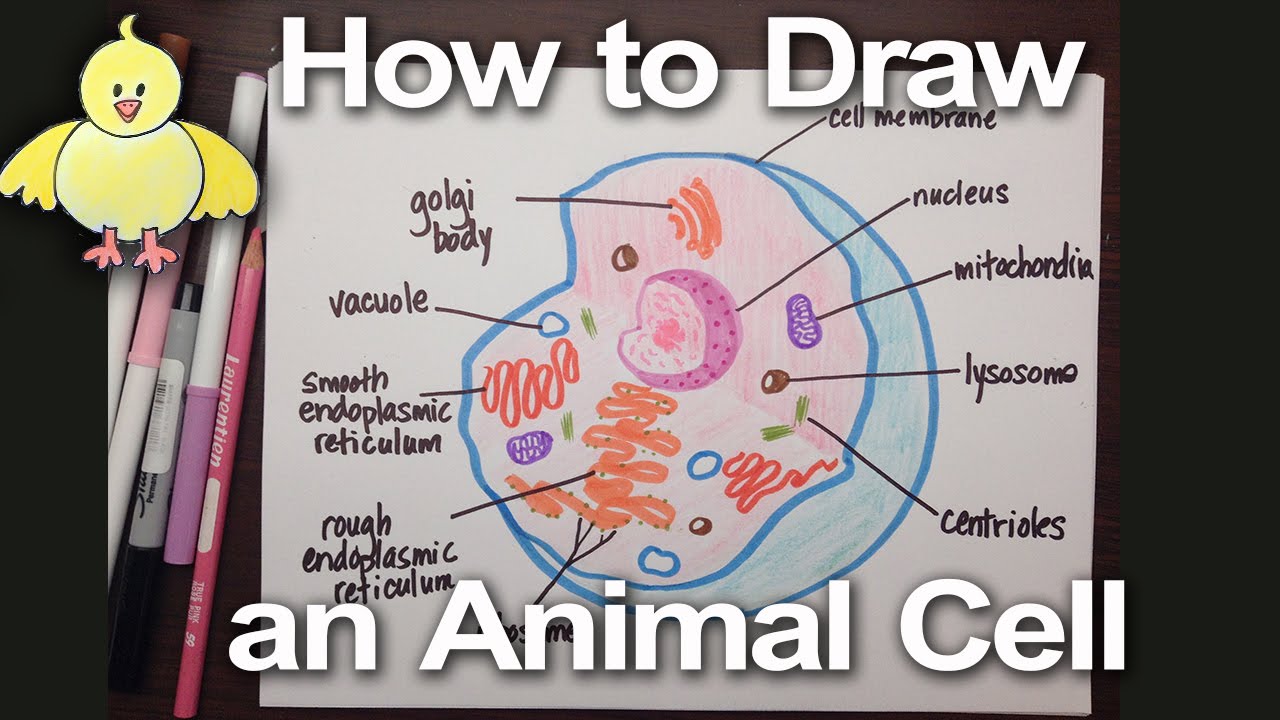
How to Draw an Animal Cell: 11 Steps (with Pictures) - wikiHow 1. Draw a simple circle or oval for the cell membrane. The cell membrane of an animal cell is not a perfect circle. You can make the circle misshapen or oblong. The important part is that it does not have any sharp edges. [1] Also know that the membrane is not a rigid cell wall like in plant cells.

Animal Cell Diagram - Tim's Printables
A Labeled Diagram of the Animal Cell and its Organelles A Labeled Diagram of the Animal Cell and its Organelles There are two types of cells - Prokaryotic and Eucaryotic. Eukaryotic cells are larger, more complex, and have evolved more recently than prokaryotes. Where, prokaryotes are just bacteria and archaea, eukaryotes are literally everything else.

Basics of Animal Cell Biology | LoveToKnow
Free Animal Cell Diagram Templates - Edrawsoft A clear design animal cell diagram template from Edraw is waiting for you in the free download version. Use it for any kinds of science coursework or group discussions. You can also adjust the diagram sizes at any time you want for more insights.
Animal Cell- Definition, Structure, Parts, Functions, Labeled ...
Printable Animal Cell Diagram - Labeled, Unlabeled, and Blank Printable animal cell diagram to help you learn the organelles in an animal cell in preparation for your test or quiz. 5th grade science and biology. Soul Candy Inter. Design (253) 376-9675 Homeschool
133,122 Plant cell Images, Stock Photos & Vectors | Shutterstock
Basic Virology, Third Edition - Academia.edu Basic Virology, Third Edition . ... As a result, the infected cell is dramatically different from the ... Download Free PDF View PDF. Journal of Virology. Virion factors targeting Daxx to overcome intrinsic immunity. 2013 • Sabrina Schreiner. Download Free PDF View PDF. Leonard Norkin Virology Molecular Biology and Pathogenesis.pdf. Mohamed Ali . Download Free PDF View …
Animal Cell - Free printable to label + Color -kidCourses.com
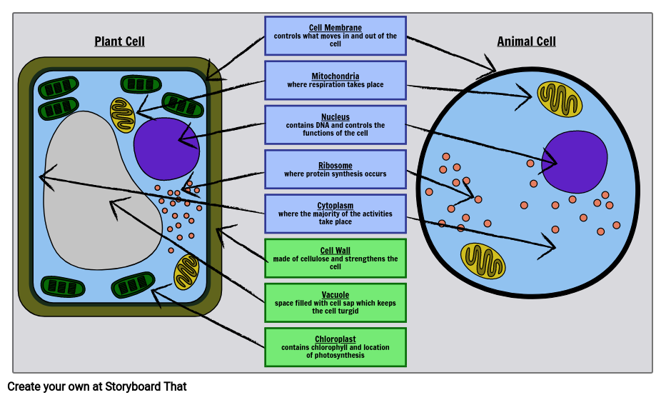
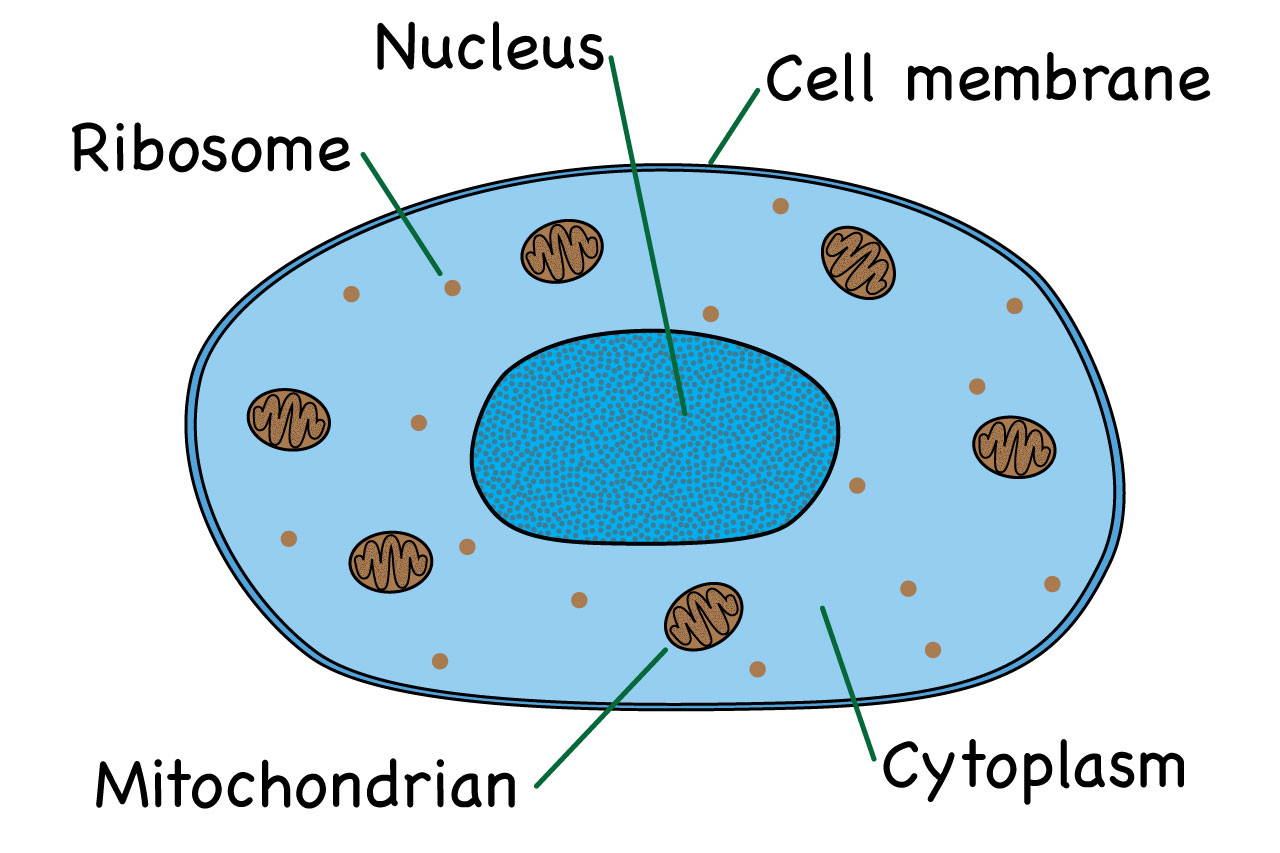
Animal Cell Labelling Activity | Basic Animal Cell Diagram - Twinkl Animal cells have four key components: Nucleus: the command centre of the cell. Cell membrane: the cell 'wall'. Cytoplasm: the liquid component, which holds water and nutrients. Mitochondria: the cell 'powerhouse', which converts food into energy for the body to use. The above video may be from a third-party source.

cell | Definition, Types, Functions, Diagram, Division ...
Animal Cell - resource - Imageshare - Benetech Basic educational diagram showing a cross section of an animal cell. Design modalities for the image include braille with and without labels, print with and without labels in greyscale, color, and texture.

Label the Animal Cell Worksheets (SB11866) | Animal cells ...
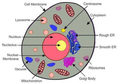
Animal Cell Diagram | Science Trends An animal cell diagram is a great way to learn and understand the many functions of an animal cell. The diagram, like the one above, will include labels of the major parts of an animal cell including the cell membrane, nucleus, ribosomes, mitochondria, vesicles, and cytosol.
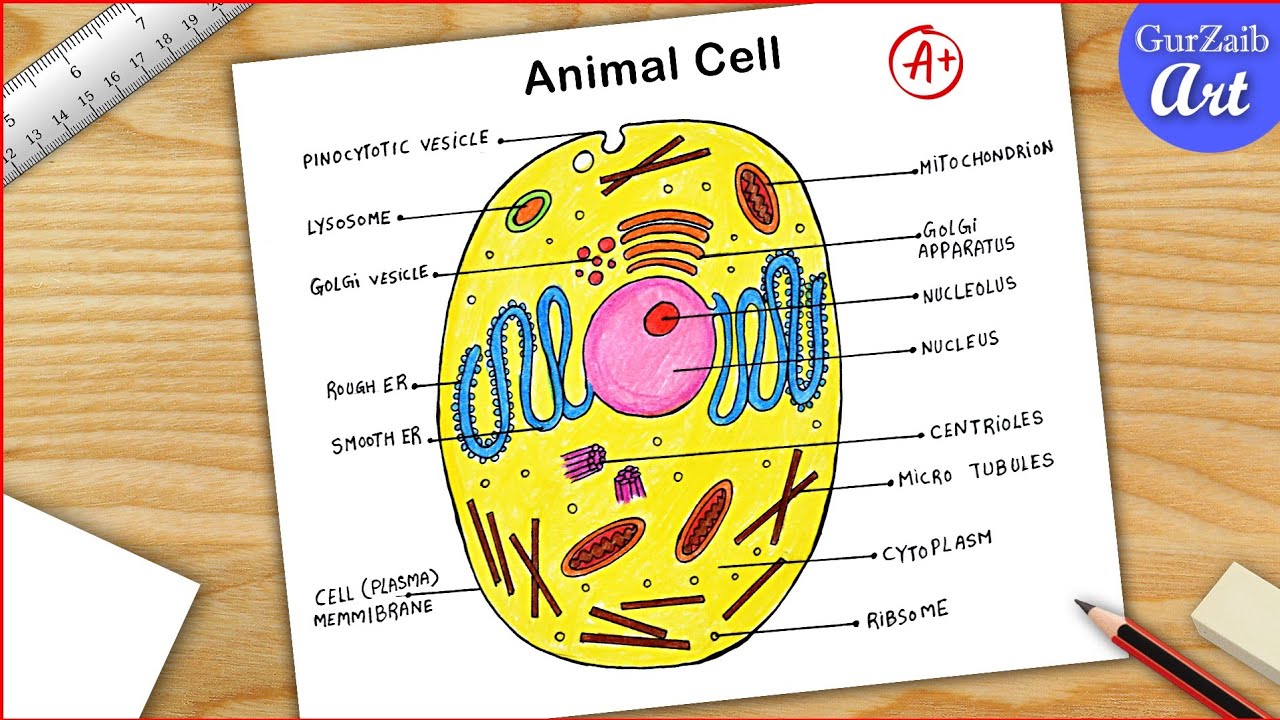
Animal Cell Diagram Drawing CBSE || easy way || labeled Science project - Step by step
Animal Cell- Definition, Structure, Parts, Functions, Labeled Diagram The largest animal cell is the ostrich egg which has a 5-inch diameter, weighing about 1.2-1.4 kg and the smallest animal cells are neurons of about 100 microns in diameter. Animal cells are smaller than the plant cells and they are generally irregular in shape taking various forms of shapes, due to lack of the cell wall.
Free Animal Cell Diagram Templates
Animal Cell - Structure, Function, Diagram and Types - BYJUS Animal Cell Types There are numerous types of animal cells, each designed to serve specific functions. The most common types of animal cells are: Skin Cells Melanocytes, keratinocytes, Merkel cells and Langerhans cells Muscle Cells Myocyte, Myosatellite cells, Tendon cells, Cardiac muscle cells Blood Cells Leukocytes, erythrocytes, platelet
Animal cell with labeled anatomic structure parts diagram outline diagram
GLYPHICONS - Basic set Basic set. Buy Basic set $ 149. Get a better price with new collections. Author: Jan Kovařík. Icons: 1060+ Version: 2.4. License: Regular license. Files: Vector files (AI, SVG, PDF, EPS) Raster files (PNG, @2x, @3x) Design trends may come and go, but simplicity and comprehensibility will never go out of style. This continuously evolving pack of icons is an investment for the future, you will ...
Plant Cell- Definition, Structure, Parts, Functions, Labeled ...
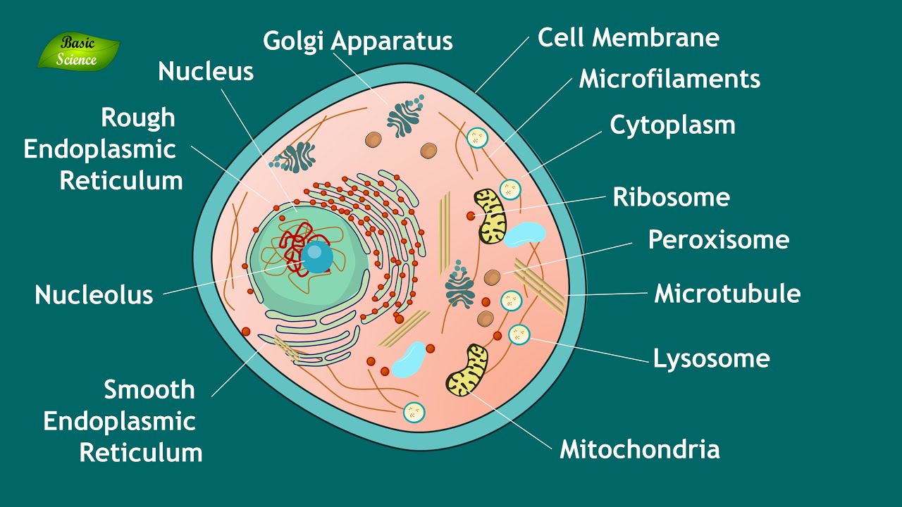
Animal Cell diagram with labels by Russell Kightley Media Labelled Animal Cell Diagram ABOVE: Animal Cell Illustration with labels showing major organelles including: nucleus nucleolus nuclear envelope nuclear pore centrioles cell membrane microtubule smooth endoplasmic reticulum rough endoplasmic reticulum ribosomes polysomes lysosome peroxisome mitochondrion Golgi ( plant cells are somewhat different).
1,654 Animal cell diagram Images, Stock Photos & Vectors ...
Guidelines for Safe Work Practices in Human and Animal ... 06.01.2012 · Regardless, animal cadavers can harbor zoonotic agents, and risk assessment to determine whether zoonotic infectious agents may be present in a cadaver, as outlined in Section 12, is critically important for establishing appropriate animal necropsy biosafety procedures. The guidelines in this section are combined biosafety best practices for both human autopsy and …
Plant Cell Structures and Functions | Let's Talk Science
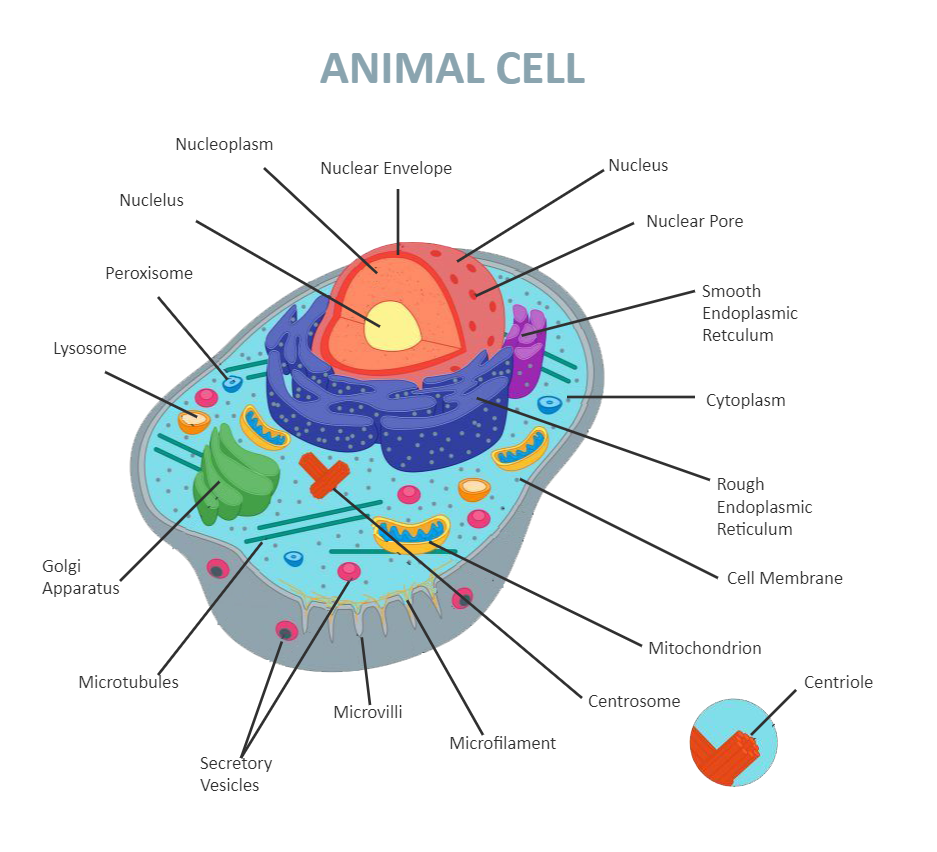
Animal Cell - The Definitive Guide | Biology Dictionary Labeled diagram of a typical animal cell Nucleus The nucleus contains all the genetic material in a cell. This genetic information is called deoxyribonucleic acid (DNA). DNA contains all the instructions for making proteins, which control all of the body's activities. Therefore, the nucleus is like the manager's office of the cell.
Animal cell diagram hi-res stock photography and images - Alamy
Plant Cell: Diagram, Types and Functions - Embibe Exams The cell is the basic structural and functional unit of life in all living organisms. The cells can be divided into two major groups - Prokaryotic and Eukaryotic. The difference between both the cells are explained below: Prokaryotic cell: Cell without a well-defined nucleus, i.e. cell of bacteria.

Bex Taylor - KS3 Animal Cell Worksheet
Eukaryotic Cell: Definition, Structure & Function (with Analogy & Diagram) The vacuole: Plant cells contain at least one large vacuole to maintain the cell's shape, while animal vacuoles are smaller in size. The centriole: Animal cells have one; plant cells don't. Chloroplasts: Plant cells have them; animal cells don't. The cell wall: Plant cells have an outer cell wall; animal cells simply have the plasma membrane.
Animal cell diagram hi-res stock photography and images - Alamy
Free Printable Plant and Animal Cells Worksheets 27.06.2022 · This animal cell diagram is labeled in cursive writing for your students who are learning about ... Plant Cell Parts Color Poster – This free plant cell parts poster is in full color to give a basic illustration of a plant cell including labels for the nucleus, chloroplast, cytoplasm , membrane, cell wall, vacuole, and mitochondrion. Learning About Plant Cells Diagram Printable …
Label Cell Parts | Plant & Animal Cell Activity | StoryboardThat
Animal and Plant Cell Worksheets - Super Teacher Worksheets This is a basic illustration of a plant cell with major parts labeled. Labels include nucleus, chloroplast, cytoplasm, membrane, cell wall, and vacuole, and mitochondrion. Use it as a poster in your classroom or have students glue it into their science notebooks. View PDF Plant Cell Vocabulary Cards
Animal Cell Structure and Function | Notes | Eukaryotic Cell | Basic Science Series
Interactive Bacteria Cell Model - CELLS alive Periplasmic Space: This cellular compartment is found only in those bacteria that have both an outer membrane and plasma membrane (e.g. Gram negative bacteria).In the space are enzymes and other proteins that help digest and move nutrients into the cell. Cell Wall: Composed of peptidoglycan (polysaccharides + protein), the cell wall maintains the overall shape of a …

Animal Cell Model Diagram Project Parts Structure Labeled ...

Animal Cells and the Membrane-Bound Nucleus
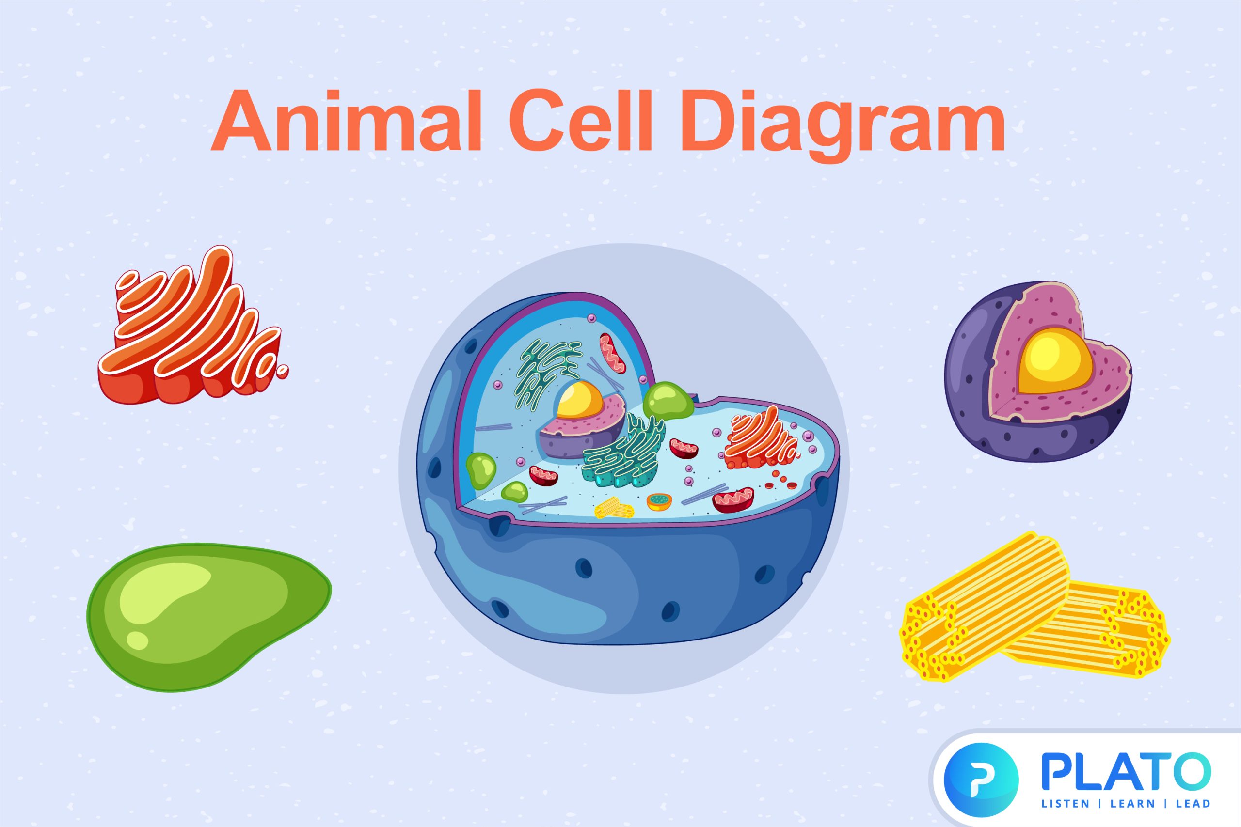
Animal Cell Diagram - Plato Online
A Well-labelled Diagram Of Animal Cell With Explanation

File:Simple diagram of animal cell (en).svg - Wikimedia Commons
Animal Cell Diagram
Animal Cell - Structure, Function, Diagram and Types

The Cell | AmoebaMike
What Is An Animal Cell? Facts, Pictures & Info For Kids ...
Animal cell label - Teaching resources

A Labeled Diagram of the Animal Cell and its Organelles ...

File:Simple diagram of animal cell (numbers).svg - Wikimedia ...
Animal cell diagram hi-res stock photography and images - Alamy
How to Draw an Animal Cell Diagram -Homework Help | DoodleDrawArt
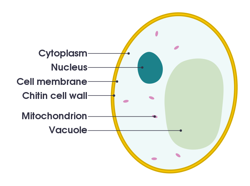
File:Simple diagram of yeast cell (en).svg - Wikipedia

Animal Cell Structure and Organelles with their functions
Plant Cell - Definition, Structure, Function, Diagram & Types
Post a Comment for "41 basic animal cell diagram with labels"