39 fluorescent labels and light microscopy
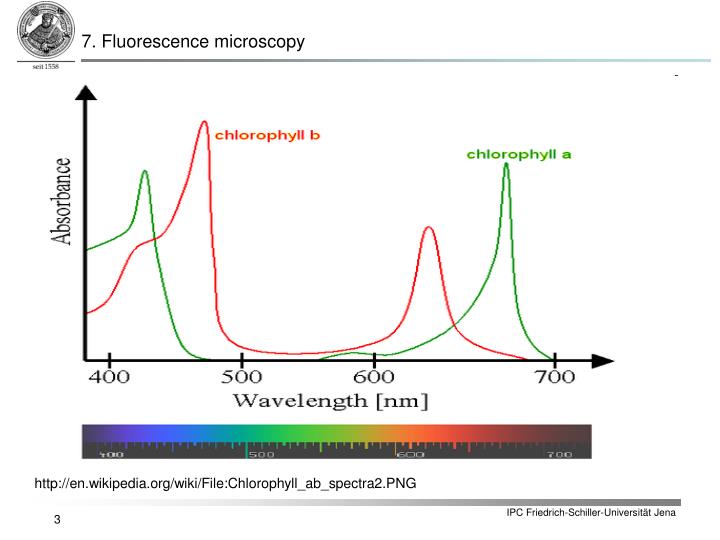
Fluorescence - Wikipedia Fluorescence is the emission of light by a substance that has absorbed light or other electromagnetic radiation.It is a form of luminescence.In most cases, the emitted light has a longer wavelength, and therefore a lower photon energy, than the absorbed radiation. A quick guide to light microscopy in cell biology - PMC Fluorescence microscopy uses fluorescent dyes (fluorophores), which are molecules that absorb one wavelength of light (the excitation wavelength) and emit a second, longer wavelength of light (the emission wavelength).
Fluorescence Microscopy - an overview | ScienceDirect Topics M. Heilemann, in Comprehensive Biophysics, 2012 Abstract. Fluorescence microscopy is a valuable toolbox to study cellular structures and dynamics spanning scales from the single molecule to the live animal. The spatial resolution that can be achieved with any light-based microscopy is however limited to about 200 nm in the imaging plane and >500 nm along the optical axis.
Fluorescent labels and light microscopy
Super resolution fluorescence microscopy - PMC Among the various microscopy techniques, fluorescence microscopy is one of the most widely used because of its two principal advantages: Specific cellular components may be observed through molecule-specific labeling, and light microscopy allows the observation of structures inside a live sample in real time. Fluorescence Microscopy vs. Light Microscopy - Medical News This means that fluorescent microscopy uses reflected rather than transmitted light. For example, a commonly used label is green fluorescent protein (GFP), which is excited with blue light and... Label-free prediction of three-dimensional fluorescence images from ... We present a label-free method for predicting three-dimensional fluorescence directly from transmitted-light images and demonstrate that it can be used to generate multi-structure, integrated...
Fluorescent labels and light microscopy. Fluorescent Labeling - What You Should Know - PromoCell Definition Fluorescent labeling is the process of binding fluorescent dyes to functional groups contained in biomolecules so that they can be visualized by fluorescence imaging (nature.com). The availability of new fluorophores has dramatically changed the possibilities for the sensitive detection of biomolecules and the analysis of their interactions. Improved fluorescent dyes are now ... A quick guide to light microscopy in cell biology Light microscopy is a key tool in modern cell biology. Light microscopy has several features that make it ideally suited for imaging biology in living cells: the resolution is well-matched to the sizes of subcellular structures, a diverse range of available fluorescent probes makes it possible to mark proteins, organelles, and other structures for imaging, and the relatively nonperturbing ... Introduction to Fluorescent Proteins | Nikon’s MicroscopyU The brightness and fluorescence emission spectrum of enhanced yellow fluorescent protein combine to make this probe an excellent candidate for multicolor imaging experiments in fluorescence microscopy. Enhanced yellow fluorescent protein is also useful for energy transfer experiments when paired with enhanced cyan fluorescent protein (ECFP) or ... Smart Microscope for Lab Routine and Research - ZEISS In the past, documenting samples with multiple fluorescent labels in your routine lab could be time consuming. To get best image quality, you needed to manually switch filters, adjust illumination intensities and exposure times and to snap each single channel image. For four different channels, this could sum up to 15 steps and clicks.
Light Microscope- Definition, Principle, Types, Parts, Labeled Diagram ... A light microscope is a biology laboratory instrument or tool, that uses visible light to detect and magnify very small objects and enlarge them. They use lenses to focus light on the specimen, magnifying it thus producing an image. The specimen is normally placed close to the microscopic lens. Basics of FRET Microscopy | Nikon’s MicroscopyU The first fluorescent protein biosensor was a calcium indicator named cameleon, constructed by sandwiching the protein calmodulin and the calcium calmodulin-binding domain of myosin light chain kinase (M13 domain) between enhanced blue and green fluorescent proteins (EBFP and EGFP). In the presence of increasing levels of intracellular calcium ... Fluorescence Imaging - Teledyne Photometrics Fluorescent molecules (known as fluorophores) are used to label samples, and fluorophores are available that emit light in virtually any color. In a fluorescent microscope, a sample is labeled with a fluorophore, and then a bright light ( excitation light) is used to illuminate the sample, which gives off fluorescence ( emission light ). Fluorescent Labelling - an overview | ScienceDirect Topics Fluorescence microscopy Fluorescent labeling methods are generally based on reactive derivatives of fluorophores that selectively bind to functional groups contained in target biomolecules and are widely used in biotechnology because of their non-destructive properties and the high sensitivity of fluorescence techniques ( Sahoo, 2012 ).
Novel Fluorescent Label Shines a Light on DNA Structure in Cancer Cells Liu and her team formulated a new label called Hoechst-Cy5 to overcome the hurdle that fluorescent dyes didn't work well on DNA or in processed clinical cancer samples. After showing that the new... Principles of Fluorescence and Fluorescence Microscopy Fluorescence Microscopy Author: Carl Zeiss Microscopy GmbH, Germany Date: May 2019 Fluorescence is the property of atoms and molecules, so called fluorophores, to absorb light at a particular wavelength and to subsequently emit light of longer wavelength. Fluorescence microscopy can be based on autofluorescence or the addition of fluorescent dyes. Fluorescence-Based Methods for the Study of Protein Localization ... Standardized fluorescent protein labeling methods for filamentous fungal proteins (Toews et al. 2004; Yang et al. 2004; Yu et al. 2004) in conjunction with fluorescence or confocal microscopy (see Chap. 1) have enabled in vivo analyses of protein dynamics. This has been achieved mainly through time-lapse acquirements over short- or long-time ... Advanced fluorescence microscopy techniques for the life sciences To facilitate observation at the single molecule level 14, a fluorescent molecule, referred to as a label or a fluorophore, is attached to the biological molecule of interest, excited using a coherent light source and the emitted fluorescence is, consequently, collected.
FLUORESCENCE MICROSCOPY/CELL BIOLOGY: The planar truth about light-sheet microscopy | Laser ...
PDF Light-Sheet Fluorescence Microscopy - UConn Health With light-sheet fluorescence microscopy (LSFM) - also known as selective plane illumination microscopy (SPIM) - a conceptually new method was introduced to fluorescence live imaging in 2004. This development by Ernst Stelzer and his group at the European Molecular Biology Laboratory (EMBL) in Heidelberg, published in Huisken et al 2004, 5
Frontiers | Applications of Light-Sheet Microscopy in Microdevices | Frontiers in Neuroanatomy
Fluorescence Microscopy - Explanation and Labelled Images Fluorescence microscopy uses a high-intensity light source that excites a fluorescent molecule called a fluorophore in the sample observed. The samples are labeled with fluorophore where they absorb the high-intensity light from the source and emit a lower energy light of longer wavelength.

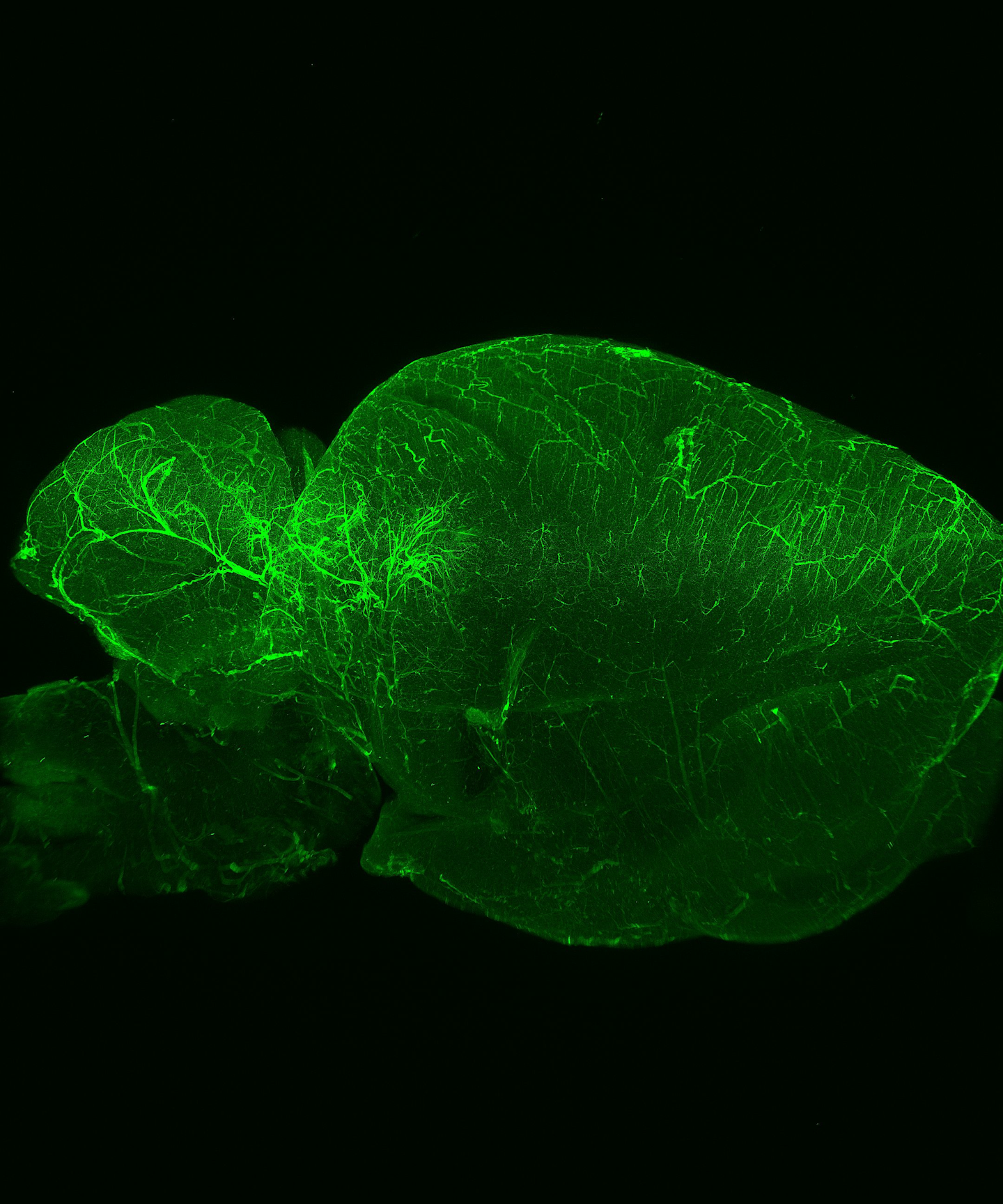
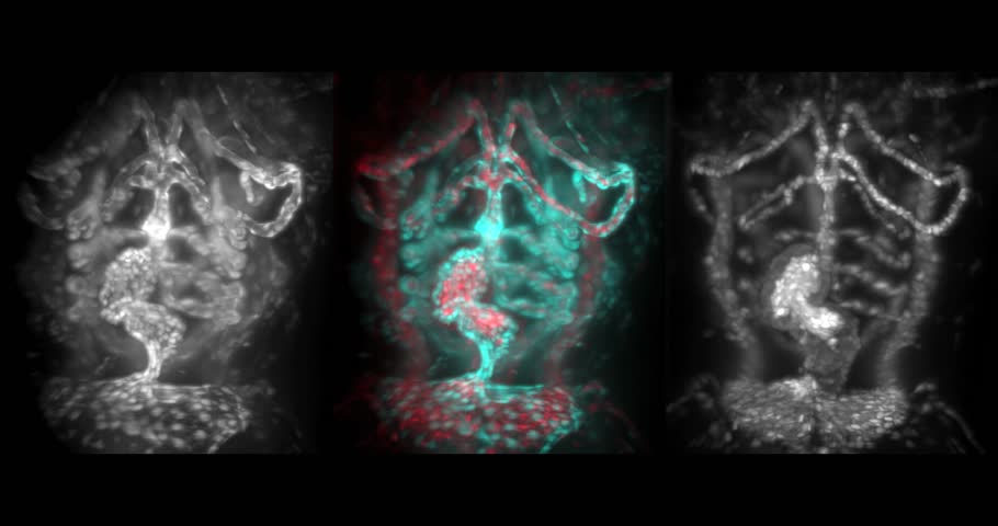
Advancing multiscale structural mapping of the brain through fluorescence imaging and analysis ...
PDF Saturated excitation microscopy for sub-diffraction-limited imaging of ... Keywords: confocal microscopy; fluorescence microscopy; saturated excitation; depth discrimination property; high resolution. Paper 130420RR received Jun. 18, 2013; revised manuscript received Oct. 28, 2013; accepted for publication Nov. 1, 2013; published ... high-image contrast. 1 4 A pinhole placed in front of a light detector is a key ...

Protein and Cellular Engineering | Department of Pharmaceutical Chemistry | UCSF
Fluorescent tag - Wikipedia Fluorescent tag. S. cerevisiae septins revealed with fluorescent microscopy utilizing fluorescent labeling. In molecular biology and biotechnology, a fluorescent tag, also known as a fluorescent label or fluorescent probe, is a molecule that is attached chemically to aid in the detection of a biomolecule such as a protein, antibody, or amino acid.
PPT - 7.1 Fluorochromes Fluorescence microscopy differentiates between two kinds of ...
Saturated excitation microscopy for sub-diffraction-limited imaging of ... Saturated excitation (SAX) microscopy offers high-depth discrimination predominantly due to nonlinearity in the fluorescence response induced by the SAX. Calculation of the optical transfer functions and the edge responses for SAX microscopy revealed the contrast improvement of high-spatial frequency components in the sample structure and the effective reduction of background signals from the ...

RFP-Booster | Chromotek
Label-free prediction of three-dimensional fluorescence images from ... We present a label-free method for predicting three-dimensional fluorescence directly from transmitted-light images and demonstrate that it can be used to generate multi-structure, integrated...

产品中心 - 成都阿帕克生物科技有限公司
Fluorescence Microscopy vs. Light Microscopy - Medical News This means that fluorescent microscopy uses reflected rather than transmitted light. For example, a commonly used label is green fluorescent protein (GFP), which is excited with blue light and...
Light Sheet Fluorescence Microscopy Stock Video Footage - 4K and HD Video Clips | Shutterstock
Super resolution fluorescence microscopy - PMC Among the various microscopy techniques, fluorescence microscopy is one of the most widely used because of its two principal advantages: Specific cellular components may be observed through molecule-specific labeling, and light microscopy allows the observation of structures inside a live sample in real time.
FLUORESCENCE MICROSCOPY: A program to help labs 'go green' | Laser Focus World

Label-free prediction of three-dimensional fluorescence images from transmitted-light microscopy ...

Label-free prediction of three-dimensional fluorescence images from transmitted-light microscopy ...
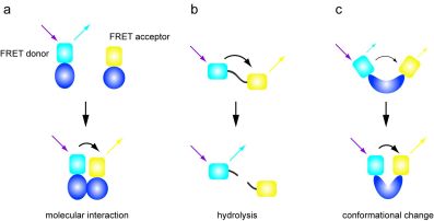
FRET and Translocation in Cell-based Imaging - 2009 - Wiley Analytical Science

Label-free prediction of three-dimensional fluorescence images from transmitted-light microscopy ...

Click Chemistry With Copper - A Biocompatible Version | Berkeley Lab

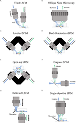
Experimental configurations of single-molecule fluorescence microscopy.... | Download Scientific ...
Post a Comment for "39 fluorescent labels and light microscopy"