39 picture of compound microscope with labels and functions
meltingclock.co › printable-free-empathyPrintable Free Empathy Worksheets Pdf Aug 20, 2021 · Developing empathy year university undergraduates language phenomenological study worksheets pdf. Discover learning games, guided lessons, and other interactive activities for children. › pmc › articlesLEARNING FROM OTHERS: CHILDREN’S CONSTRUCTION OF CONCEPTS - PMC There is a rich empirical literature showing that children’s initial, non-linguistic concepts are affected by the labels they hear (e.g., “a bird”, “a wug”). Labels can be considered a form of testimony, but I consider them separately, because there is a large literature on lexicalization effects, and they raise a distinct set of issues.
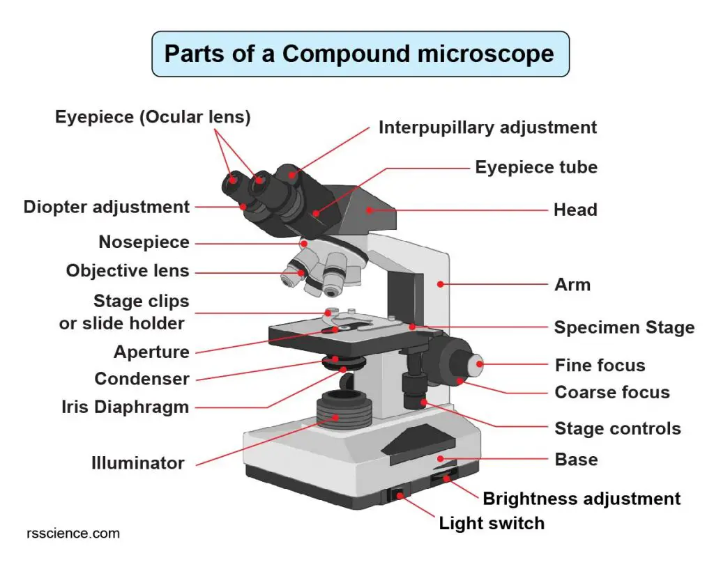
Parts of a microscope with functions and labeled diagram Head - This is also known as the body. It carries the optical parts in the upper part of the microscope. Base - It acts as microscopes support. It also carries microscopic illuminators. Arms - This is the part connecting the base and to the head and the eyepiece tube to the base of the microscope.
Picture of compound microscope with labels and functions
› knowledge-pathwayAn Intro to Immunofluorescence: Staining & Processing for ... It is filtered light that excites a fluorochrome, a fluorescing compound, that is attached to an antibody. The most frequently use fluorochromes are fluorescein, or FITC , rhodamine, phycoerythrin (PE), Dylight, Texas Red. There are many Cy labels, Cy2, Cy3 are quite popular, and many Alexa Fluor fluorescent labels as well. Compound Microscope: Parts of Compound Microscope It is a U-shaped structure and supports the entire weight of the compound microscope. 2. Pillar. It is a vertical projection. This stands by resting on the base and supports the stage. 3. Arm. The entire microscope is handled by a strong and curved structure known as the arm. 4. Label the microscope - Science Learning Hub In this interactive, you can label the different parts of a microscope. Use this with the Microscope parts activity to help students identify and label the main parts of a microscope and then describe their functions. Drag and drop the text labels onto the microscope diagram. If you want to redo an answer, click on the box and the answer will ...
Picture of compound microscope with labels and functions. Microscope Parts and Functions With Labeled Diagram and ... Body tube (Head): The body tube connects the eyepiece to the objective lenses. Arm: The arm connects the body tube to the base of the microscope. Coarse adjustment: Brings the specimen into general focus. Fine adjustment: Fine tunes the focus and increases the detail of the specimen. Nosepiece: A rotating turret that houses the objective lenses ... Simple Microscope - Parts, Functions, Diagram and ... Compound microscope - It comes with more than one lens and provides better magnification than the simple microscope. A compound microscope is also called a bright field microscope. It can provide magnification by up to 1,000 times. Stereo microscope/dissecting microscope - It can magnify objects by up to 300 times. It is used to visualize ... 16 Parts of a Compound Microscope: Diagrams and Video ... In compound microscopes with two eye pieces there are prisms contained in the body that will also split the beam of light to enable you to view the image through both eye pieces. 2. Arm. The arm of the microscope is another structural piece. The arm connects the base of the microscope to the head/body of the microscope. Compound Microscope - Types, Parts, Diagram, Functions and ... Eyepiece/ocular lens - It is the part of the microscope that is looked through at the top. It comes with a magnification ranging between 5x and 30x. Image 3: The head connects the eyepiece to the objective lens. Picture Source: microscope.com. Head (monocular/binocular) - It is the structural support of the microscope.
Compound Microscope Labeled Diagram - Quizlet QUESTION. The total magnification of a specimen being viewed with a 10X ocular lens and a 40X objective lens is. 15 answers. QUESTION. a mosquito beats its wings up and down 600 times per second, which you hear as a very annoying 600 Hz sound. if the air outside is 20 C, how far would a sound wave travel between wing beats. 2 answers. Compound Microscope: Definition, Diagram, Parts, Uses ... A compound microscope is defined as. A microscope with a high resolution and uses two sets of lenses providing a 2-dimensional image of the sample. The term compound refers to the usage of more than one lens in the microscope. Also, the compound microscope is one of the types of optical microscopes. The other type of optical microscope is a ... Compound Microscope Parts, Function, & Diagram | What is a ... Learn the compound light microscope's parts and functions by viewing a compound microscope diagram. Also, read about the uses of a compound microscope. Updated: 11/04/2021 What is a Compound Microscope? - New York Microscope Company A compound microscope is an instrument that is used to view magnified images of small specimens on a glass slide. It can achieve higher levels of magnification than stereo or other low power microscopes and reduce chromatic aberration. It achieves this through the use of two or more lenses in the objective and the eyepiece.
Compound Microscope Parts - Labeled Diagram and their ... The term "compound" refers to the microscope having more than one lens. Basically, compound microscopes generate magnified images through an aligned pair of the objective lens and the ocular lens. In contrast, "simple microscopes" have only one convex lens and function more like glass magnifiers. [In this figure] Two "antique ... Labelled Diagram of Compound Microscope - Biology Discussion The below mentioned article provides a labelled diagram of compound microscope. Part # 1. The Stand: The stand is made up of a heavy foot which carries a curved inclinable limb or arm bearing the body tube. The foot is generally horse shoe-shaped structure (Fig. 2) which rests on table top or any other surface on which the microscope in kept. drdollah.com › laboratory-information-system-lisLaboratory Information System (LIS) | HEALTHCARE SERVICE DELIVERY Sep 06, 2014 · Full Blood Picture. Blood from the specimen is smeared on to a glass slide, stained and viewed under the microscope. It is important that bar code labels are attached to the slide to ensure continuity of matching specimen to the right patient. Histopathology and Cytology Examination Compound Microscope- Definition, Labeled Diagram ... The optical microscope often referred to as the light microscope, is a type of microscope that uses visible light and a system of lenses to magnify images of small subjects. There are two basic types of optical microscopes: Simple microscopes. Compound microscopes. The term "compound" in compound microscopes refers to the microscope having ...
Compound Microscope Parts – Labeled Diagram and their Functions - Rs' Science
Compound Microscope - Diagram (Parts labelled), Principle ... A compound microscope: Is used to view samples that are not visible to the naked eye. Uses two types of lenses - Objective and ocular lenses. Has a higher level of magnification - Typically up to 2000x. Is used in hospitals and forensic labs by scientists, biologists and researchers to study microorganisms.
Compound Microscope Clipart | Free Images at Clker.com - vector clip art online, royalty free ...
worksheetstudent.comWorksheet Student - Worksheet Website for Students May 01, 2022 · The worksheets include first grade appropriate reading passages and related questions. 1st grade reading comprehension worksheets printable pdf today the number of words that can...

Labeling the Parts of the Microscopes including Free Printouts
Parts of a Compound Microscope and Their Functions Compound microscope mechanical parts include base or foot, pillar, arm, inclination joint, stage, clips, diaphragm, body tube, nose piece, coarse adjustment knob and fine adjustment knob. Base: It's the horseshoe-shaped base structure. All of the other components of the compound microscope are supported by it.

31 Drag The Label To The Appropriate Part Of The Microscope. - Labels Design Ideas 2020
Diagram of a Compound Microscope - Biology Discussion 1. It is noted first that which objective lens is in use on the microscope. 2. Stage micrometer is positioned in such a way that it is in the field of view. 3. The eyepiece is rotated so that the two scales, the eyepiece or ocular scale and the stage micrometer scale, are parallel. 4.

Compound Light Microscope Parts And Functions Worksheet | Decoratingspecial.com
Microscope Types (with labeled diagrams) and Functions A compound microscope: Is used to view samples that are not visible to the naked eye. Uses two types of lenses - Objective and ocular lenses. Has a higher level of magnification - Typically up to 2000x. Is used in hospitals and forensic labs by scientists, biologists and researchers to study micro organisms. Compound microscope labeled diagram.

All Saints Online: Microscope Part Functions
Parts of Microscope, Function, Names & Labeled Diagram ... Condenser. The condenser is to focus the light, which passes from the microscopic illuminator to the specimen. This condenser is located just below the diaphragm. This diaphragm is one of the important parts of the compound microscope which will help to get an accurate and sharp image. The condenser has a magnification power of 400X and above.

Biology 2201
Labeling the Parts of the Microscope | Microscope activity ... Description A collection of microscope diagrams and worksheets for science class. Download them all in one convenient PDF, and select the version that's best for your classroom. This PDF contains the following: 1. Parts of a Microscope Diagram - Color 2. Parts of a Microscope Diagram - Black and White 3.

Parts of a microscope with functions and labeled diagram
› curriculum-linksCurriculum Links & Videos | Oak Meadow Homeschool 76: Functions of (-x), Functions of the Other Angle, Trigonometric Identities (1), Rules of the Game Instructional Video: Even and Odd Functions (Trig) Instructional Video: Complementary Angles (Trig) Instructional Video: Verifying Trig Identities. Oak Meadow Lesson 20 (Textbook Lessons 77-80) 77: Binomial Expansions (1)
Post a Comment for "39 picture of compound microscope with labels and functions"